
With lichen planopilaris, quiet inflammation can still cost follicles.
Burning, tenderness and perifollicular scale may accompany patchy loss, but symptoms can be subtle. Once a follicle is replaced by scar tissue, regrowth from that opening is unlikely.
Lichen planopilaris is an immune-mediated scarring alopecia.
Inflammation targets the upper follicle and can progressively destroy its regenerative structures. Patchy loss commonly occurs on the crown or vertex, while variants can involve the frontal hairline.
Treatment aims to control activity, ease symptoms and preserve follicles that remain. It cannot reliably restore follicles already destroyed.
Openings can disappear
Loss of visible follicular openings is a key clue that distinguishes scarring from many non-scarring disorders.
Symptoms vary
Itching, burning, pain and tenderness may be prominent, mild or absent.
The edge is most informative
Redness and scale around remaining hairs at an active margin can guide biopsy.
Course is hard to predict
Activity may wax and wane, so repeat examination is often more useful than one symptom snapshot.
Biopsy the question, not simply the emptiest spot.
An established smooth center may contain too little active disease to classify. Dermatologists often select a symptomatic or inflamed margin where follicles are still present.
Clinic-pathology correlation matters because several scarring alopecias can overlap in appearance.
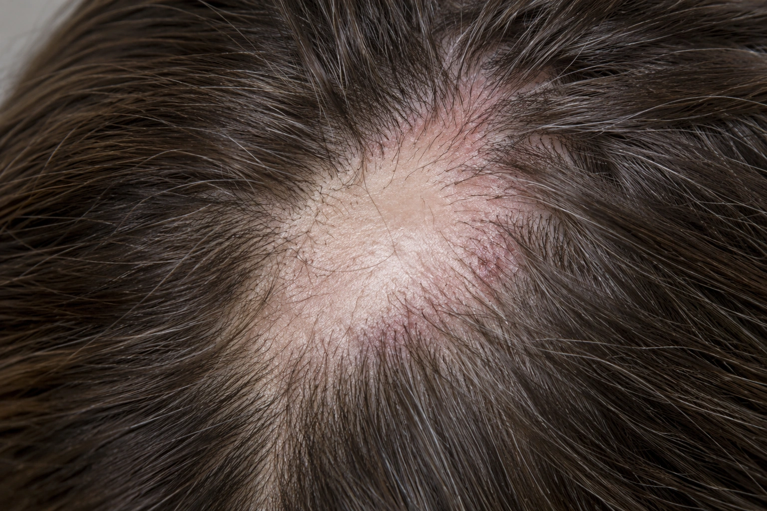
Look for the combination of loss, inflammation and disappearing openings.
The pattern warrants timely medical assessment even when symptoms are tolerable.
- Irregular patchy hair loss
- Perifollicular scale or redness
- Burning, itching or tenderness
- Tufted or lonely remaining hairs
- Loss of follicular openings
- Frontal fibrosing alopecia
- Discoid lupus erythematosus
- Central centrifugal cicatricial alopecia
- Folliculitis decalvans
- Alopecia areata or traction
Dermatology diagnosis should not wait for extensive visible scarring.
A clinical trichologist can identify concerning changes and monitor photographs, but biopsy and prescription management belong with an appropriate medical clinician.
- 01 · Map symptoms and marginsDocument burning, pain, itch, scale and progression across scalp zones.
- 02 · Use trichoscopyLook for perifollicular scale, erythema, casts and absent openings.
- 03 · Select an active biopsy siteA dermatologist may sample the margin rather than a fully scarred center.
- 04 · Monitor disease activityCombine symptoms, examination and photographs instead of relying on shedding alone.
When the story needs medical attention.
Hair loss may be one clue within a larger health issue. Seek timely evaluation when any of these apply.
- Rapidly expanding scar-like loss
- Increasing pain or burning
- Pustules, crusting or drainage
- Eyebrow loss with frontal recession
- Mucosal symptoms or widespread rash
- Medication side effects
Control inflammation early and review whether the margins are still active.
Treatment is individualized to extent, symptoms, progression, comorbidities and risk tolerance.
Topical or injected corticosteroids
May be used for limited active areas under medical supervision.
Anti-inflammatory or immune therapy
Hydroxychloroquine, tetracyclines and other agents may be considered by a dermatologist.
Choice, monitoring and evidence vary; response is not guaranteed.Gentle scalp care
Reduce irritation and treat coexisting dermatitis without substituting cosmetic care for disease control.
Wait for durable stability
Transplantation or cosmetic procedures are considered cautiously after prolonged inactivity because recurrence can affect results.
Disease control is judged over repeated visits.
Confirm suspected scarring
Arrange dermatology assessment when openings are disappearing or symptoms are active.
Check activity
Compare symptoms, scale, redness and expansion at the margins.
Refine maintenance
Balance stability, treatment risk and quality of life with the treating clinician.
Lichen Planopilaris
Recognize the scalp symptoms and follicular changes that make prompt dermatology assessment important.
Read next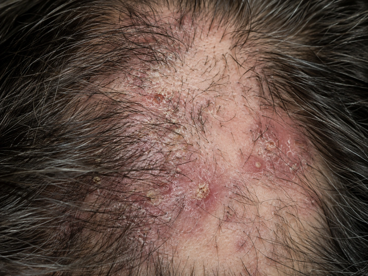

Clear answers to the next questions.
Can LPP hair grow back?
Hair cannot usually regrow from follicles already replaced by scar tissue; preserving remaining follicles is the priority.
Is LPP contagious?
No. It is an inflammatory immune-mediated condition.
Can it stop on its own?
Activity can fluctuate or become quiet, but waiting without assessment risks avoidable follicle loss.
Does scalp pain measure severity?
Not reliably. Some active disease causes few symptoms, so examination matters.
Why might I need a biopsy?
It can help distinguish LPP from other scarring or inflammatory disorders and guide treatment.
Can I have a hair transplant?
Only selected, durably stable cases may be considered; recurrence and limited graft survival remain concerns.