
Lichen planopilaris
Patchy hair loss with burning, itching, tenderness, redness, or scale around individual hairs can signal active inflammation that may permanently destroy follicles.
Educational guide · A dermatologist should diagnose and medically manage suspected scarring alopecia

What is lichen planopilaris?
Lichen planopilaris—often shortened to LPP—is an uncommon inflammatory disease in which immune activity damages scalp follicles and replaces them with scar tissue.
- It most often creates irregular, progressive patches.
- The exact cause is unknown, and it is not contagious.
- Success means controlling activity and preventing further loss.
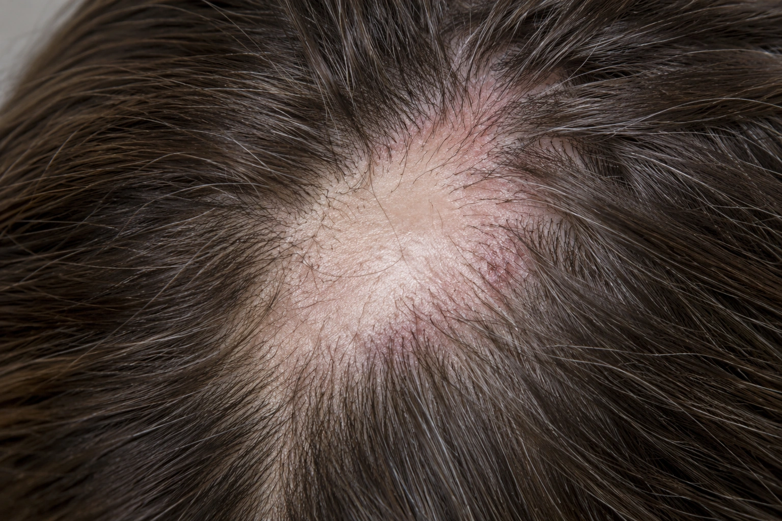
The border tells a different story than the center.
A quiet, smooth center may represent damage that has already occurred. Redness and scale around remaining follicles at the edge may indicate current activity.
Redness and perifollicular scale
Fine scale can form a collar around individual hair shafts.
Smooth, pale, or shiny skin
Normal follicular openings may be reduced or absent.
Itching, burning, pain, or tenderness
Symptoms vary and may be mild—or absent—even when disease is active.
Lichen planopilaris can appear in several recognized forms.
The distribution matters because related conditions can affect the frontal hairline, eyebrows, scalp, and other hair-bearing areas differently.
Irregular scalp patches
Multifocal areas may occur across the scalp, often with inflammation at the margin and smooth scarring within established patches.
Band-like frontal recession
The frontal and temporal hairline gradually recedes; eyebrow and body-hair loss may accompany it.
A characteristic triad
Scarring scalp loss occurs with non-scarring loss in the armpits or groin and rough follicular bumps on the body.
Inflammation targets structures needed for future growth.
 EDUCATIONAL FOLLICLE CROSS-SECTION
Follicle opening present
A viable follicle supports a growing hair and remains visible at the scalp surface.
Inflammation develops
Immune cells concentrate around the upper follicle, with redness and scale at the surface.
Scar tissue replaces it
After destructive injury, the opening disappears and that follicle cannot produce another hair.
Anti-inflammatory care cannot restore a follicle that is already scarred. It is intended to suppress active disease, relieve symptoms, and protect follicles that have not been destroyed.
A biopsy is most useful when it samples an active edge.
Clinical pattern, trichoscopy, and sometimes pathology work together. A completely scarred, inactive center may contain fewer diagnostic clues.
History and symptoms
Document onset, progression, itch, burning, tenderness, hairline or eyebrow change, health history, and treatments.
Trichoscopy
Magnification may show perifollicular redness and tubular scale, white areas or dots, and missing follicular openings.
Dermatologist-led biopsy
A sample from a clinically active margin can distinguish LPP from other inflammatory and scarring alopecias.
A verified trichologist can document patterns, symptoms, and change, then coordinate referral. Diagnosis, biopsy, prescription therapy, and management of inflammatory disease require an appropriately licensed healthcare professional.
Not every patchy or inflamed scalp is LPP.
Discoid lupus
Another immune-mediated scarring alopecia with overlapping clinical signs but different pathology.
CCCA
Commonly begins at the central crown and expands outward; presentation and risk profile differ.
Folliculitis decalvans
Pustules, crusting, drainage, and tufted hairs point toward a neutrophilic scarring process.
Stabilization is the measure of success.
There is no single treatment that works for everyone. A dermatologist individualizes care according to activity, extent, symptoms, health history, and treatment risk.
Local anti-inflammatory care
Prescription topical or injected medicines may be used for limited active areas.
Selection follows medical evaluationSystemic treatment
Progressive or extensive disease may require dermatologist-supervised oral therapy and monitoring.
Benefits and risks varyGentle scalp support
Reduce friction and irritating practices while controlling symptoms; supportive care does not replace treatment.
Comfort—not a cureMeasure activity
Repeat photographs, symptoms, trichoscopy, and examination help show whether progression has stopped.
Track change consistentlyHair transplantation or surgery is considered only in carefully selected, long-stable disease. Recurrence and reduced graft survival remain possible; no restorative procedure guarantees results.
The aim is no new permanent loss.
Disease activity can be difficult to judge from a single day. Compare symptoms and standardized scalp findings over time.
No expanding patches
Existing areas remain unchanged and no new scarred zones appear.
Less inflammation
Redness, scale, tenderness, itching, or burning improve.
Remaining hair is preserved
Loss at the active margins slows or stops.
Follow-up remains consistent
Quiet symptoms do not automatically prove that disease is inactive.
New scalp symptoms with patchy loss deserve prompt evaluation.
Arrange dermatology assessment for burning, pain, tenderness, persistent itching, redness or scale around hairs, an enlarging irregular patch, eyebrow loss, or smooth shiny skin where follicular openings seem absent.
Find a verified trichologist →Lichen planopilaris, clearly explained.
Is lichen planopilaris contagious?+
No. Lichen planopilaris is an inflammatory immune-mediated condition and cannot be passed from one person to another.
Will hair grow back in affected areas?+
Hair cannot regrow from follicles that have been destroyed and replaced by scar tissue. Treatment focuses on controlling active inflammation and protecting follicles that remain viable.
Does lichen planopilaris always itch or burn?+
No. Itching, burning, pain, and tenderness can occur, but some people have few symptoms. Visible change and close scalp examination remain important.
Why is a scalp biopsy sometimes needed?+
Several scarring alopecias can look alike. A dermatologist may biopsy an active edge—where redness or scale surrounds remaining hairs—to identify the inflammatory pattern and guide treatment.
Is frontal fibrosing alopecia the same condition?+
Frontal fibrosing alopecia is generally considered part of the lichen planopilaris spectrum, but its band-like frontal pattern and frequent eyebrow involvement give it a distinct clinical presentation.
How do I know whether treatment is working?+
The goal is stability: fewer symptoms, less redness and scale, no enlargement of scarred areas, and no new patches. Standardized photographs and repeat clinical examinations help track change.
Medical review notes
This guide preserves the existing Trichology.com foundation—immune-mediated inflammation, patchy scarring, recognized variants, symptoms, trichoscopy, biopsy, treatment categories, prognosis, and support—while prioritizing early consumer recognition.
Medical disclaimer: This content is educational and is not a diagnosis or substitute for care from a qualified health professional.
Place LPP within the scarring-alopecia spectrum.
Compare the broader category and closely related patterns before drawing conclusions from appearance alone.