
The Androgen-Hair Axis
Follow DHT synthesis and androgen-receptor signaling within the follicle.

 William Gaunitz, WTSClinical Trichologist · Expert Reviewer
Inside this review
Use these questions to navigate the complete physician-authored review below.
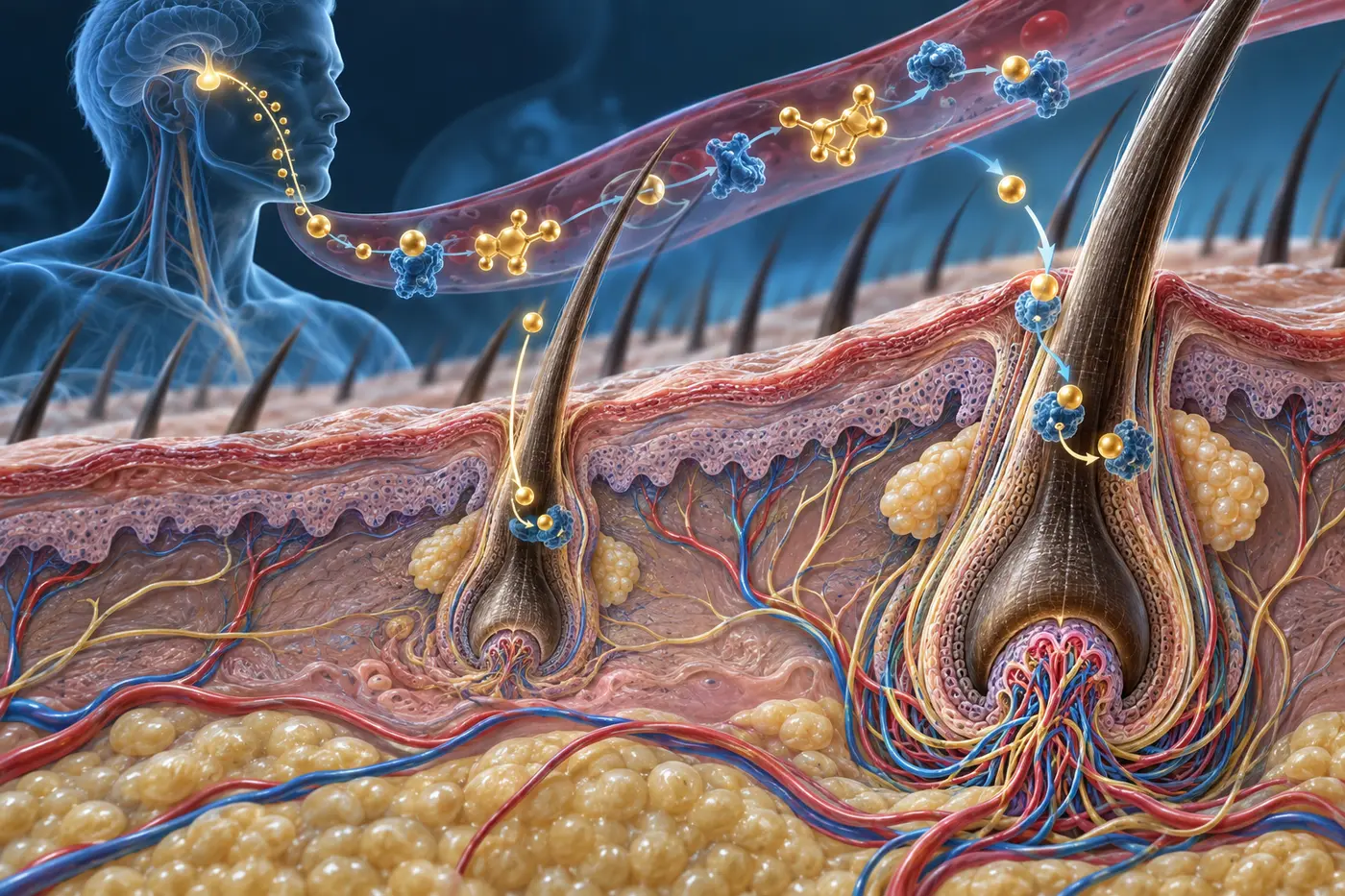
Follow the conversion of testosterone to DHT and the role of 5-alpha reductase.
Read this section →Explore androgen receptors and the local follicular response to androgen signaling.
Read this section →Complete production article and literature cited.
The Androgen-Hair Axis
How DHT, Androgen Receptors, and Dermal Papilla Signaling Drive Follicular
Miniaturization
Key Takeaways
- Androgens are the primary hormonal drivers of pattern hair loss. Dihydrotestosterone (DHT) binds to androgen receptors in the dermal papilla, triggering a cascade of signaling changes that progressively shorten the anagen phase.
- Genetic sensitivity determines susceptibility. The density and activity of androgen receptors within follicular tissue, not circulating androgen levels, determines how severely a follicle responds to DHT.
- Follicular miniaturization is a gradual, reversible-to-irreversible spectrum. Early miniaturization reflects altered signaling, not permanent follicle loss. Advanced stages involve structural changes that limit regenerative capacity.
- Androgen excess affects women differently than men. Female pattern hair loss involves distinct hormonal mechanisms, but androgenic effects only drive miniaturization in follicles with an underlying genetic sensitivity to DHT.
- Effective treatment requires targeting the hormonal pathway. Blocking DHT production or receptor binding can slow or halt miniaturization, but timing and follicle viability determine the outcome.
Introduction
Androgenetic alopecia (AGA) is the most prevalent form of hair loss worldwide, affecting approximately 50% of men by the age of 50 and a significant proportion of women across all age groups. Despite its familiarity, the biological mechanisms driving this condition are frequently oversimplified in clinical settings, often reduced to the single explanation of “genetic baldness.”
The reality is substantially more complex. Androgenetic alopecia is not the result of hair follicles spontaneously disappearing. Instead, it reflects a progressive transformation driven by the interaction between androgens, specifically dihydrotestosterone (DHT), and genetically sensitized follicles. This interaction unfolds across multiple levels of biology, from hormone biosynthesis and receptor binding through to downstream alterations in the dermal papilla, growth factor secretion, and hair cycle timing.
Understanding this pathway in mechanistic detail is essential for modern trichology practice. It explains why treatments such as finasteride and dutasteride are effective, why some patients respond to treatment while others do not, and why intervening early in the miniaturization process is critical for preserving follicular function.
This article examines the androgen-hair axis from first principles: how DHT is produced, how it interacts with follicular tissue, what biological changes follow that interaction, and what this means for clinical evaluation and management.
DHT Biosynthesis and the Role of 5-Alpha Reductase
Testosterone is the primary circulating androgen in both men and women, though at significantly different concentrations. While testosterone itself has moderate androgenic activity in peripheral tissues, its conversion to dihydrotestosterone (DHT) amplifies androgenic signaling substantially. This conversion is mediated by the enzyme 5-alpha reductase (5-AR), which exists in two functionally relevant isoforms: type 1 and type 2.

Figure 1. Conversion of testosterone to dihydrotestosterone (DHT) by 5-alpha reductase.⁹
Type 2 5-alpha reductase is the dominant isoform within the dermal papilla of scalp follicles and is responsible for the majority of intrafollicular DHT production. DHT binds to the androgen receptor with approximately five times greater affinity than testosterone and dissociates from it more slowly, resulting in a far more potent and sustained androgenic signal within susceptible follicles.³
Critically, DHT exerts its effects locally. Serum androgen concentrations do not necessarily predict follicular androgen exposure. A patient may have entirely normal circulating testosterone levels while still experiencing significant intrafollicular DHT activity, driven by elevated local 5-AR expression or amplified receptor sensitivity.
This distinction has important clinical implications. Measuring serum androgens is insufficient for assessing follicular androgen burden. Conversely, it also means that interventions targeting 5-AR activity, regardless of systemic androgen levels, can meaningfully reduce the DHT signal reaching the follicle.
Androgen Receptor Biology in the Hair Follicle
The androgen receptor (AR) is a nuclear receptor protein that, upon binding to DHT, undergoes conformational changes that allow it to translocate into the nucleus and regulate gene transcription.⁴ In the context of hair follicles, AR expression is concentrated in the dermal papilla, the mesenchymal signaling center of the follicle, rather than in the epithelial matrix cells that directly produce the hair shaft.

Figure 2. Hair follicle anatomy showing sebaceous gland and dermal papilla.¹⁰
This anatomical specificity is central to understanding how DHT mediates its effects. The dermal papilla does not produce hair directly but instead controls hair growth by secreting paracrine signals that regulate the behavior of adjacent stem cells and matrix cells. When androgen receptor signaling within the dermal papilla is activated by DHT, the pattern of these paracrine signals changes in ways that are hostile to sustained hair growth.
The number of androgen receptors expressed within follicular tissue varies between individuals and between different body regions. Scalp follicles in androgen-sensitive areas, including the frontal hairline, vertex, and crown, express significantly higher androgen receptor density than occipital follicles, which remain largely resistant to androgenic miniaturization. This regional difference in receptor expression is genetically determined and explains the characteristic distribution patterns of androgenetic alopecia.
Importantly, androgen receptor gene polymorphisms that influence receptor sensitivity have been identified as major genetic risk factors for androgenetic alopecia.⁵ This means that susceptibility to AGA is not determined solely by the amount of DHT produced but also by how aggressively the receptor responds to that signal.
Dermal Papilla Signaling Changes Driven by DHT
When DHT activates androgen receptor signaling in the dermal papilla, several downstream consequences alter the follicular microenvironment in ways that impair hair growth.
Among the most well-characterized effects is the increased production of dickkopf-related protein 1 (DKK-1), a potent inhibitor of Wnt/beta-catenin signaling.⁵ As discussed in the context of follicular stem cells, Wnt signaling is the primary activator of stem cells in the bulge region. By upregulating DKK-1, DHT effectively suppresses the molecular trigger required to initiate a new growth phase. The result is prolonged resting periods and shortened anagen cycles.
DHT also reduces the dermal papilla’s production of pro-growth paracrine factors, including insulin-like growth factor-1 (IGF-1) and vascular endothelial growth factor (VEGF).⁷ IGF-1 promotes matrix cell proliferation and hair shaft elongation, while VEGF supports the perifollicular microvascular network that delivers oxygen and nutrients. The simultaneous reduction of both factors creates a microenvironment that is nutrient-poor, poorly oxygenated, and metabolically compromised.
In parallel, DHT-activated dermal papilla cells upregulate transforming growth factor-beta 1 (TGF-beta 1), a cytokine that promotes premature entry into the catagen phase. TGF-beta 1 triggers apoptosis in matrix cells and accelerates follicular regression, cutting short the productive period of each growth cycle.
The cumulative effect of these signaling changes, including reduced Wnt activation, loss of IGF-1 and VEGF support, and elevated TGF-beta 1, produces successively shorter and weaker growth cycles with each passing hair cycle. The follicle gradually miniaturizes.
The Miniaturization Process: From Terminal to Vellus
Follicular miniaturization is the defining histological feature of androgenetic alopecia.⁸ It describes the progressive reduction in follicle size, hair shaft diameter, and anagen duration that occurs as androgen receptor signaling reshapes follicular biology over successive hair cycles.
In the earliest stages of miniaturization, follicles produce hair shafts of near-normal diameter but with progressively shorter anagen phases. Because the hair cycle is shorter, each hair reaches a lesser total length before shedding. The scalp hair appears thinner and less dense but individual hairs are not yet dramatically reduced in caliber.
As the process advances, the dermal papilla itself shrinks. A smaller dermal papilla communicates with a proportionally smaller population of matrix cells, producing a hair shaft
with reduced diameter and pigmentation. These intermediate hairs, sometimes referred to as miniaturized terminal hairs, are finer than normal but remain pigmented.
In fully advanced miniaturization, the follicle produces vellus-caliber hairs: unpigmented, fine, and short. At this stage, the dermal papilla has become substantially reduced in volume, and the total anagen duration may be as brief as weeks rather than years. The scalp appears visibly thinned or bald in affected areas.
A critical distinction must be drawn between follicular miniaturization and follicular loss. In most cases of androgenetic alopecia, including advanced stages, follicles remain present within the scalp. They are not destroyed but rather persistently suppressed. This distinction is what makes early intervention therapeutically meaningful. Follicles that have miniaturized but not yet converted to fibrotic scar tissue retain the potential to produce thicker hair if the androgenic signal is reduced or interrupted.
Androgenetic Alopecia in Women: A Distinct Hormonal Context
Female pattern hair loss (FPHL) shares the fundamental mechanism of androgenetic alopecia, with androgen receptor activation driving follicular miniaturization, but occurs within a different hormonal environment that produces clinically distinct features.
In women, testosterone is produced in smaller quantities by the ovaries and adrenal glands. Estrogens, which predominate in the female hormonal milieu, generally exert protective effects on hair growth by prolonging the anagen phase. This estrogen-androgen balance helps explain why female pattern hair loss typically presents later and with a different distribution than male AGA, favoring diffuse thinning over the crown rather than a receding hairline.
However, several conditions can disturb this balance and accelerate androgenic effects on scalp follicles. Polycystic ovary syndrome (PCOS), elevated circulating androgens from adrenal sources, and relative androgen excess following menopause all represent states in which follicular androgen receptor activation is amplified. In postmenopausal women, the decline in estrogen production removes a key counterbalance to androgen activity, often accelerating hair thinning.
Importantly, many women with FPHL present with normal serum androgen levels. In these cases, the issue lies not with systemic androgen excess but with local follicular androgen sensitivity, a finding consistent with the known variability in androgen receptor expression across individuals.
This distinction has direct implications for evaluation. Measuring androgens is a worthwhile component of the workup for female hair loss, particularly when other signs of androgen excess are present. However, a normal androgen profile does not exclude androgenetic alopecia as the diagnosis.
Treatment Implications: Targeting the Androgen-Hair Axis
Understanding the androgen-hair axis in mechanistic detail clarifies how and why specific treatments are effective, and why they must be deployed strategically.
5-Alpha Reductase Inhibitors
Finasteride and dutasteride reduce intrafollicular DHT by inhibiting the 5-AR enzyme. Finasteride selectively inhibits type 2 5-AR, while dutasteride inhibits both type 1 and type 2 isoforms, achieving greater reductions in DHT levels. By reducing the androgenic signal reaching the dermal papilla, these agents slow or halt miniaturization and, in some patients, allow partial restoration of follicular size over time.
Their effectiveness depends critically on follicular viability at the time of treatment. Follicles that are still producing miniaturized but non-scarred hair retain regenerative potential. Follicles that have been replaced by fibrotic tissue do not respond. This biological reality underscores the clinical value of early detection and intervention.
Androgen Receptor Antagonists
Spironolactone and, more recently, bicalutamide are used in female patients to block androgen receptor activation directly. By competing with DHT for receptor binding, these agents reduce the downstream signaling consequences within the dermal papilla. They are particularly relevant for women with documented androgen excess or pronounced follicular androgen sensitivity.
Combination Approaches
Because miniaturization reflects a convergence of androgenic signaling, reduced growth factor support, and impaired microvascular function, treatment strategies that address multiple aspects of this process simultaneously tend to produce superior outcomes. Combining androgen pathway intervention with agents that support Wnt signaling, improve scalp vascularity, or reduce perifollicular inflammation may provide additive benefit, particularly in patients with moderate-to-advanced miniaturization.
Implications for Trichology Practice
A working understanding of the androgen-hair axis allows trichologists to move beyond generic assessments toward mechanistically grounded evaluations. Several practical principles follow from the biology described in this article.
First, androgen receptor sensitivity, not serum androgen levels alone, determines follicular vulnerability. A thorough evaluation should consider both hormonal status and the clinical pattern of hair loss, as follicular miniaturization in the characteristic androgen-sensitive distribution provides direct evidence of receptor-mediated activity even in the presence of normal laboratory values.
Second, the reversibility of miniaturization is time-dependent. Follicles that have recently entered the miniaturization process retain greater regenerative capacity than those that have undergone years of progressive shrinkage. Identifying and addressing androgenic activity early expands the window of treatment effectiveness.
Third, the androgen-hair axis intersects with multiple other biological systems described in prior articles in this series. Iron availability affects the metabolic capacity of follicles under androgenic stress. Immune privilege, which can be disrupted by perifollicular inflammation, is further compromised in follicles that have been functionally weakened by androgen signaling. Scalp microvascular function, already reduced by DHT’s suppression of VEGF, determines how effectively therapeutic agents and nutrients reach the affected follicle.
These intersections mean that optimizing androgen pathway management alongside broader follicular support is likely to produce better outcomes than isolated pharmacological intervention.
Conclusion
Androgenetic alopecia is not simply the consequence of inherited baldness. It is the clinical expression of a specific molecular pathway in which DHT, acting through androgen receptors concentrated in the dermal papilla, progressively dismantles the signaling architecture that supports healthy hair cycling.
This pathway, encompassing 5-alpha reductase activity, androgen receptor activation, and downstream alterations in Wnt, IGF-1, VEGF, and TGF-beta 1 signaling, explains the characteristic progression from shortened anagen cycles to follicular miniaturization and eventual hair loss. It also explains why interventions targeting this pathway can halt or partially reverse the process, provided that viable follicles remain.
For trichology practice, the androgen-hair axis represents one of the most clearly defined targets in hair loss medicine. Accurate evaluation of its activity, through pattern recognition, hormonal assessment, and an understanding of the interplay with iron metabolism, immune privilege, stem cell signaling, and scalp microenvironment, provides the foundation for rational, evidence-based management of the most common form of hair loss encountered in clinical practice.
References
¹ Stenn KS, Paus R. Controls of hair follicle cycling. Physiol Rev. 2001;81(1):449–494. https://doi.org/10.1152/physrev.2001.81.1.449
² Tobin DJ. Biochemistry of human skin—our brain on the outside. Chem Soc Rev. 2006;35(1):52–67. https://doi.org/10.1039/B505793K
³ Randall VA. Androgens and hair growth. Dermatol Ther. 2008;21(5):314–328. https://doi.org/10.1111/j.1529-8019.2008.00214.x
⁴ Inui S, Itami S. Androgen actions on the human hair follicle: perspectives. Exp Dermatol. 2013;22(3):168–171. https://doi.org/10.1111/exd.12024
⁵ Trueb RM. Molecular mechanisms of androgenetic alopecia. Exp Gerontol. 2002;37(8–9):981–990. https://doi.org/10.1016/S0531-5565(02)00093-1
⁶ Hibino T, Nishiyama T. Role of TGF-β2 in the human hair cycle. J Dermatol Sci. 2004;35(1):9–18. https://doi.org/10.1016/j.jdermsci.2003.12.003
⁷ Yano K, Brown LF, Detmar M. Control of hair growth and follicle size by VEGF-mediated angiogenesis. J Clin Invest. 2001;107(4):409–417. https://doi.org/10.1172/JCI11317
⁸ Sinclair R, Torkamani N, Jones L. Androgenetic alopecia: new insights into the pathogenesis and mechanism of hair loss. F1000Res. 2015;4:585. https://doi.org/10.12688/f1000research.6401.1
⁹ National Center for Biotechnology Information. Conversion of testosterone to dihydrotestosterone by 5-alpha reductase. NCBI. 2020. Available at: https://www.researchgate.net/figure/The-conversion-of-dihydrotestosterone-from-testosteron e-by-5-alpha-Reductase-The-figure_fig1_350155740
¹⁰ TeachMeAnatomy. Hair follicle anatomy showing sebaceous gland and dermal papilla. Available at: https://teachmeanatomy.info/the-basics/ultrastructure/hair-follicle/